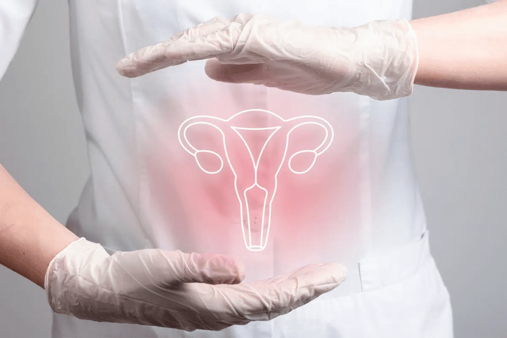
How is Biopsy Done in Cervical Cancer? – Dr. Neelu Prasad Explains
Biopsy is a crucial test used to confirm the diagnosis of cervical cancer. Here’s how it is done:

What is a Cervical Biopsy?
A cervical biopsy is a procedure where a small piece of tissue is taken from the cervix (the lower part of the uterus) to be examined under a microscope for abnormal or cancerous cells.
🩺 When is it Done?
A biopsy is usually done when:
- Pap smear shows abnormal cells
- HPV test is positive for high-risk types
- Visible lesions or suspicious areas are seen during a colposcopy
🛠️ Types of Cervical Biopsy Procedures:
1. Punch Biopsy
🔹 A small tool is used to pinch off tiny samples of cervical tissue
🔹 Usually done during colposcopy
🔹 No stitches needed
2. Endocervical Curettage (ECC)
🔹 A small instrument called a curet is used to scrape cells from inside the cervical canal
🔹 Helps detect cancer higher up in the cervix
3. Cone Biopsy (Conization)
🔹 A cone-shaped section of abnormal tissue is removed from the cervix
🔹 Done using LEEP (Loop Electrosurgical Excision Procedure) or cold knife
🔹 Requires anesthesia and may be done in an operation theater
🔹 Both diagnostic and therapeutic
💬 Dr. Neelu Prasad’s Advice:
“If your Pap smear or HPV test is abnormal, don’t panic. A cervical biopsy is a safe and essential step for accurate diagnosis. Early detection means early treatment and better outcomes.”
🩹 What to Expect After the Procedure:
- Mild cramping or spotting for a few days
- Avoid tampons and intercourse for about a week
- Report any heavy bleeding, fever, or foul discharge to your doctor
🧬 Biopsy helps detect:
- Precancerous changes (CIN)
- Carcinoma in situ
- Invasive cervical cancer


